AI Veterinary Image Analyst: Species-First Imaging Review
2026-08-28
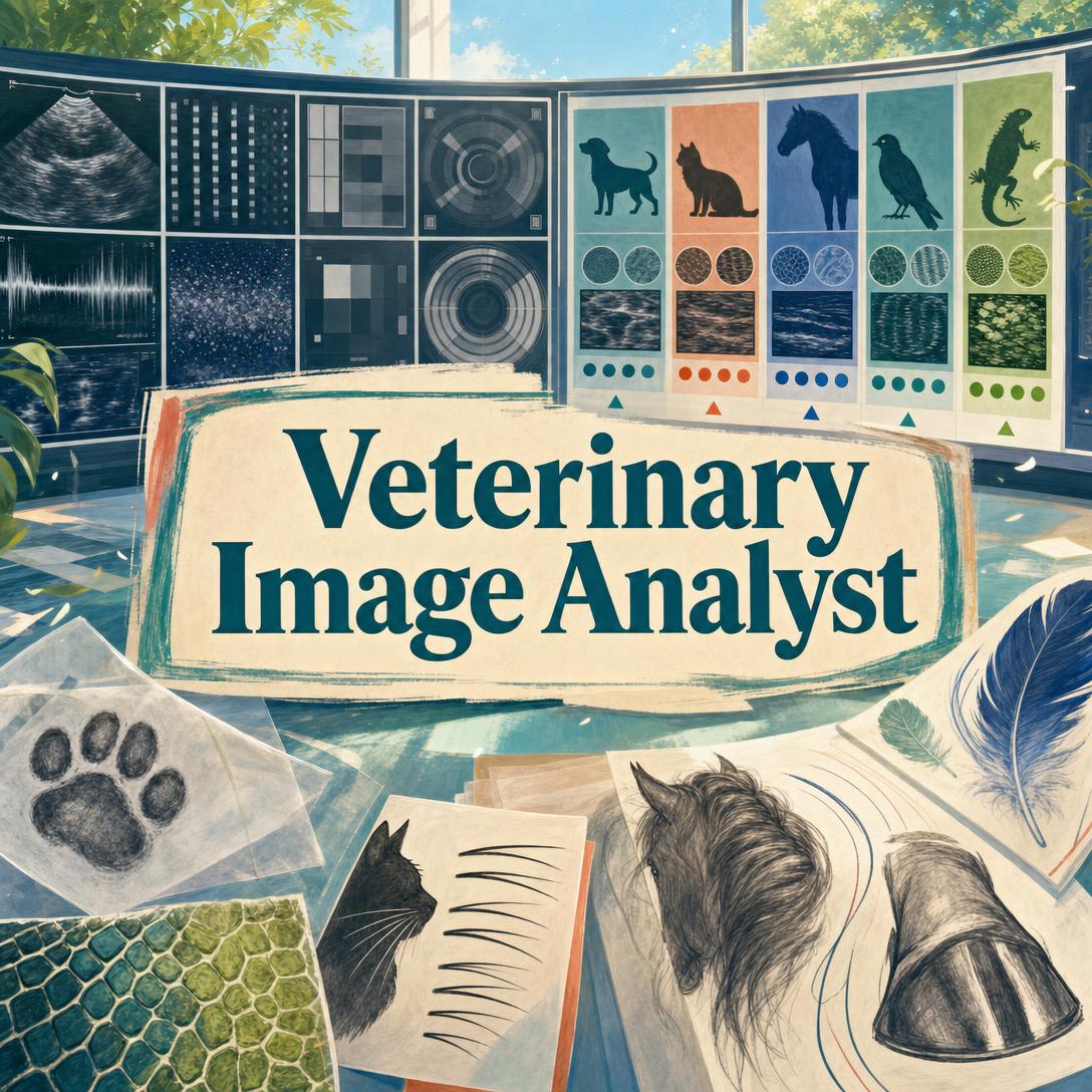
Veterinary Image Analyst helps you read veterinary images with species-first, systematic observational analysis — from canine thoracic radiographs and feline dental films to equine distal-limb studies, avian whole-body views, and cytology slides. While specialist teleradiology can be slow, costly, or simply unavailable after hours, this AI provides a structured survey of what is visible, ranked differential considerations, and a clear urgency level without claiming a diagnosis.
✅ Multi-species fluency across dogs, cats, horses, livestock, birds, reptiles, and small exotic mammals ✅ All major modalities — radiography, dental films, ultrasound, CT/MRI, cytology, endoscopy, and clinical photos ✅ Observation separated from interpretation, with honest limits when image quality is poor ✅ Urgency calibration that routes findings toward emergency care, same-week workup, or routine follow-up
To understand why that structure matters, it helps to look at how veterinary images are actually read today — and where the bottlenecks sit for clinics, students, and pet owners.
Quick Answer: What Is Veterinary Image Analyst?
Veterinary Image Analyst is a veterinary imaging specialist that reviews radiographs, ultrasound, CT, MRI, and clinical photos across all species to produce systematic, species-first observational analysis. It does not diagnose, prescribe, or give a prognosis.
Key capabilities:
- Species and breed identification before any anatomical framework is applied
- Full-image survey using Roentgen signs (size, shape, number, location, opacity/echogenicity, margin)
- Ranked differential considerations weighted by species, breed, age, and geography
- Urgency levels with specialist-type routing (emergency, surgery, internal medicine, dentistry, ophthalmology, radiology)
- Image-quality gating — poor positioning, expiration, rotation, or motion are flagged before over-reading
Why Veterinary Images Are So Easy to Misread
Diagnostic imaging is now a core part of companion-animal and large-animal practice. The global veterinary imaging market was valued at about $2.33 billion in 2025, and radiography remains the workhorse modality in most clinics. Demand is rising. Access to someone who can actually interpret those images at a specialist level is not keeping up.
Board-certified veterinary radiologists are concentrated in referral centers. General practitioners, technicians, and students are often left to read films in real time — nights, weekends, and in the field — without a second set of eyes. Pet owners encounter a different version of the same gap: a phone photo of a lump, a screenshot of a clinic monitor, or a copy of a report they do not fully understand.
Cost and geography make the gap worse.
52% of U.S. pet owners — skipped needed veterinary care in the past year, according to a PetSmart Charities–Gallup survey reported by the AVMA. Among those who declined recommended care or skipped annual visits, 71% cited finances.
Diagnostic procedures — were the most commonly declined services at 22%, ahead of preventive care and elective surgery.
~21 million households — live in areas with the least access to veterinary services.
>60% above general inflation — veterinary service prices have outpaced the broader consumer price index over two decades.
But getting a reliable read of a veterinary image is still frustratingly difficult:
- Species is not a footnote. A “large heart” on a Greyhound, a brachycephalic thorax, an avian coelom with almost no fat contrast, and a chelonian shell study are not interchangeable problems. Human or even single-species norms produce false pathology.
- One view is not a study. Thoracic staging needs three views. Orthopedic questions need orthogonals. Expiratory films mimic disease. Rotation mimics mediastinal shift. Sedation enlarges the spleen and the cardiac silhouette.
- Coded AI is not a radiology report. Commercial radiograph tools can flag normal vs. abnormal findings at high volume, but published comparisons show they are stronger at confirming normality than at catching abnormalities, and they do not generate differential diagnoses.
- Pet-owner photos and clinic-screen snapshots are now common — and easy to over-interpret or dismiss without a quality gate.
A 2025 Frontiers in Veterinary Science study compared a widely used small-animal radiograph AI with 11 board-certified veterinary radiologists across 50 canine and feline studies. The software matched the best radiologist on accuracy and was more specific, but less sensitive, and it did not provide differentials. The authors’ conclusion is the one clinics already feel: AI is most useful as a complement, not a replacement.
The American College of Veterinary Radiology (ACVR) and ECVDI take the same position: AI in diagnostic imaging should be used with a qualified veterinary professional in the loop, with transparent methods and ongoing validation. AAHA reporting notes that imaging is already one of the top two AI use cases among veterinary staff who use these tools (39.2% of staff in that survey), even as dataset transparency and third-party validation remain uneven.
This is exactly what Veterinary Image Analyst was built for: a species-first observational read that a GP, student, technician, or pet owner can use before or alongside formal specialist review — not instead of a veterinarian.
Why Veterinary Image Analyst
Veterinary Image Analyst is a standalone imaging specialist. It starts every case with species and breed, surveys the entire image before zooming in, and keeps what is seen separate from what it might mean. Confidence is calibrated to the evidence. Image limitations are stated, not smoothed over.
That is a different job from a binary “normal/abnormal” overlay on a canine or feline radiograph.
| Traditional Approach | Veterinary Image Analyst |
|---|---|
| Wait for teleradiology, or read films alone after hours | Immediate systematic observational report with urgency |
| Small-animal radiograph AI that codes findings as normal or abnormal | Multi-species, multi-modality analysis with ranked differentials |
| Human or single-species reference values applied by habit | Species-first anatomy, breed-specific context, age-appropriate norms |
| Easy to miss incidentals while chasing the presenting complaint | Full-image survey before focal interpretation |
| Pet owners left with a photo and no language for the vet visit | Plain-language pairing of technical terms, plus what to tell the veterinarian |
Species first, always
A Labrador elbow, a rabbit arcade, a horse’s lateromedial foot, and a parrot VD whole-body film do not share a template. The analyst identifies species (and breed, when visible or stated) before applying any framework. Breed-weighted differentials — GDV risk in deep-chested dogs, IVDD in chondrodystrophic breeds, tooth resorption in cats, continuously growing teeth in rabbits — inform ranking. They never override what is on the image.
Systematic survey, then focal findings
Thoracic films are read extrathoracic structures → airway → bone → cardiac silhouette (with breed-aware vertebral heart scale context when relevant) → diaphragm → pleura → pulmonary fields → vessels → hila → mediastinum. Abdominal films start with serosal detail. Musculoskeletal studies cover cortex, medulla, periosteum, joint space, and growth plates. Dental films use modified Triadan numbering. Cytology starts with sample quality, not a guess at the cell line.
Observation is not a diagnosis
Findings are described with Roentgen signs. Differentials are ranked and hedged: “characteristic of,” “consistent with,” “could represent.” Urgency is stated in time language — emergent, within 24–48 hours, within 1–2 weeks, or routine — with the specialist type that would typically see the case (DACVECC, DACVS, DACVIM, DACVR, DAVDC, DACVO, and so on). Treatment, dosing, and prognosis are out of scope, by design.
Example prompts:
"Lateral and VD thorax, 9-year-old Cavalier King Charles Spaniel, coughing at night. Walk the cardiac silhouette and pulmonary vessels before you talk about the lung fields."
"This is a horse, left front foot, lateromedial and DPr-PaDiO. I need founder distance and rotation discussed only if the image actually supports it."
"Phone photo of a mass on my 7-year-old cat’s side. Tell me what you can see, what you cannot see, and what I should ask my vet."
How Veterinary Image Analysis Works
You do not configure protocols or pick a species model. You upload the image, add whatever history you have, and the AI analyst runs a quality gate, then a full systematic read.
Step 1: Identify the patient and the study State species, breed, age, sex, and the clinical question if you know them. If the image makes species obvious, analysis can start immediately. If it does not — a cropped coelomic radiograph, a close-up of scales, an unlabeled monitor photo — you will be asked before anatomy is applied. Wrong species invalidates the rest of the report.
"Canine, 8-year-old MN Labrador, 34 kg, intermittent right forelimb lameness for 3 months. Right elbow lateral and CrCd."
Step 2: Pass the image-quality gate Positioning, exposure, motion, rotation, respiration phase, and whether the correct views are present are assessed first. An expiratory thorax that mimics an interstitial pattern is called out as such. A single lateral abdomen is treated as incomplete, not definitive. If the image is only partly diagnostic, you still get what can be said — plus the specific retake that would finish the study.
Step 3: Survey the whole image The entire field is read with a modality- and species-appropriate checklist before any focal lesion is interpreted. Incidentals are listed. Nipple shadows, skin folds, Mach bands, and superimposed collars are treated as artifacts when that is what they are.
Step 4: Findings, differentials, and urgency Notable observations come with clinical context, breed and age weighting, and a ranked differential list. Urgency is explicit. A suspected GDV or tension pneumothorax is not buried under hedging. A stable, likely incidental spondylosis in a geriatric dog is not escalated into an emergency.
Step 5: Ask only the questions that change the read Two to four follow-ups appear when they would actually refine the assessment — duration, travel history in a blastomycosis belt, whether contralateral films exist, vaccination and heartworm status, or a missing orthogonal view. If the image and history already suffice, the report does not stall on a questionnaire.
Try Veterinary Image Analyst free — no credit card required.
Veterinary Imaging Use Cases
📊 After-Hours Thoracic Radiographs in General Practice
Scenario: A 10-year-old mixed-breed dog presents at 9 p.m. with tachypnea. The GP takes a three-view thorax. The teleradiology queue will not turn around until morning.
Traditional Approach: The veterinarian reads the films alone, often under time pressure, and either refers empirically or waits overnight for a specialist report. Subtle pulmonary nodules, a small pneumothorax, or an expiratory technique pitfall are easy to miss.
Veterinary Image Analyst: A structured thoracic survey is available in the same visit — cardiac silhouette and pulmonary vessels first, then fields, pleura, and mediastinum — with an urgency call and a note on whether the study is diagnostically adequate.
- Orthogonal and three-view logic is enforced instead of over-reading a single film
- Respiration phase and rotation are treated as technical variables, not disease
- Output is written so it can be handed to a radiologist, internist, or ER clinician without being rewritten
📱 Phone Photos of Skin Lesions and Monitor Screens
Scenario: A pet owner photographs a rapidly changing mass on a cat’s flank, or a technician snaps a phone picture of the ultrasound screen during an AFAST because the PACS export is delayed. Both need a first-pass read on a phone, in the car or in the treatment bay.
Traditional Approach: Owners wait for an appointment they may delay for cost reasons. Staff wait for a better export. Meanwhile, small-animal clinic visits have fallen about 4.6% year over year, and skipped diagnostics are already common.
Veterinary Image Analyst: The same quality gate applies to a phone photo. If the image cannot support a lesion description, you are told why and how to reshoot (lighting, hair, scale, orthogonal). If it can, you get morphology, distribution, and a plain-language version of what to tell the veterinarian — not a home diagnosis.
- Works on web, iOS, and Android with the same analysis depth
- Pet-owner language is paired with the technical term on first use
- If you also need day-to-day feeding, behavior, or home-care planning around the finding, Pet Care Advisor can take the non-imaging side of that conversation
🐴 Equine and Large-Animal Field Imaging
Scenario: A sport-horse veterinarian is in a barn aisle with lateromedial and 60° proximal-distal views of a front foot, asking about rotation and sole depth before a show. A food-animal practitioner has a reproductive ultrasound on a cow and a question about what the image actually shows.
Traditional Approach: Equine distal-limb and large-animal studies are under-served by small-animal radiograph AI. Referral to a DACVR or equine sports-medicine specialist may be hours away. Pre-purchase films get a quick “looks fine” in poor light.
Veterinary Image Analyst: Equine, bovine, small-ruminant, camelid, and porcine studies are treated as their own anatomical problems — navicular region, laminitis geometry, distal-limb opacities, reproductive ultrasound windows — not as scaled-up dogs.
- Field radiographs and phone-captured monitors are quality-checked before measurements are discussed
- Pre-purchase and lameness contexts change how incidentals are framed, without inflating them
- For herd-level nutrition, housing, and production questions around the same animals, Farm & Agriculture Advisor is the operational counterpart, not a substitute for the image read
🧬 Cytology, Impression Smears, and Slide Photos
Scenario: A technician photographs an FNA slide or an ear-cytology smear and wants a structured look at quality, cell populations, and whether organisms are even in focus before the sample is discarded.
Traditional Approach: In-house cytology is often a glance at 10×. Poorly stained, ruptured, or non-representative slides still get a tentative label. Sending out every smear adds cost and a day.
Veterinary Image Analyst: Sample quality is the first line. Background, inflammatory vs. tissue cells, nuclear features, and organisms are described; criteria of malignancy are discussed only when the cells support it. Confidence stays low when stain or cellularity is inadequate.
- Staining and representativeness are called out instead of forced interpretation
- Ear cytology, skin scrapings, and fluid sediments use the same disciplined sequence
- When the question is microscopy technique or publication-quality description across non-veterinary slides, Microscopy Image Analyst covers that adjacent workflow
Frequently Asked Questions
Is Veterinary Image Analyst free?
Yes. A free tier includes core analysis with monthly usage limits. Paid plans increase capacity — Plus at $20/month, Premium at $50/month, Pro at $100/month, and higher tiers for heavier clinic or developer use. No credit card is required to start. Usage resets each billing month, with no daily caps.
Does this replace a veterinary radiologist or my veterinarian?
No. ACVR and ECVDI guidance is that imaging AI should be used with a qualified veterinary professional in the loop. Veterinary Image Analyst produces observational analysis, differential considerations, and urgency — not a diagnosis, treatment plan, or prognosis. Published work on commercial radiograph AI found it complementary to specialists, stronger at confirming normal findings than at detecting every abnormality, and silent on differentials. Treat this output as a structured second look to bring to a veterinarian.
Can it read ultrasound, CT, MRI, dental films, and cytology — or only X-rays?
All of those, plus clinical photographs, endoscopic stills, fundic images, contrast studies, and lab-report screenshots (extracting stated values, not re-reading imaging that is not in view). Small-animal radiograph AI products are typically trained on canine and feline X-rays. This specialist is built for multi-modality work and for species that never appear in those datasets — horses, birds, reptiles, rabbits, ferrets, and production animals.
How is this different from teleradiology or clinic radiograph AI?
Teleradiology remains the medicolegal specialist report. Clinic radiograph AI is often a coded normal/abnormal overlay on small-animal films. This imaging specialist sits in between: faster than a queued DACVR read, broader than a single-modality classifier, and explicit about uncertainty. It will tell you when the film is non-diagnostic. It will not invent a breed-specific vertebral heart scale or an OFA score that was not measured.
Does Veterinary Image Analyst work on mobile?
Yes. Web, iOS, and Android have full feature parity, including speech-to-text for hands-free history while you are holding a patient or a plate. Phone photos of lesions, wounds, oral cavities, and clinic monitors are accepted, then quality-gated. A blurry, off-angle, or heavily compressed snapshot is not dressed up as a complete study.
Is my imaging data used to train public AI models?
No. Data is encrypted in transit and at rest, is not sold to advertisers, and is not used to train public models. That matters in a field where dataset opacity is already a documented concern for veterinary imaging AI.
Conclusion
Veterinary images fail in predictable ways: the wrong species framework, a single non-orthogonal view, an expiratory thorax, a coded AI flag with no differential, and a pet owner staring at a photo they cannot translate into a vet visit. Specialist review is the right standard — and it is not always available the hour the film comes off the plate.
Veterinary Image Analyst gives you a species-first, whole-image observational report with ranked considerations and a clock on next steps. Use it to prepare a referral, to sanity-check an after-hours thorax, or to walk into the clinic with a precise question instead of a worried guess. Then let a licensed veterinarian make the clinical decisions.
Try Veterinary Image Analyst now. Explore more at Jenova.
For Developers: Veterinary Image Analyst is available programmatically via the Jenova API — integrate species-first veterinary image analysis into your application with a single API call. Full documentation →